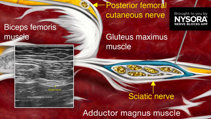
New video! Sciatic Block Anatomy Explained – NYSORA’s 3D Anatomy videos

Popliteal Block & Catheter: Crash Course with Dr. Hadzic
The popliteal block is one of the most useful regional anesthesia procedures ever. Using the all-new NYSORA Compendium of Regional Anesthesia, let’s review this nerve block. When it comes to the popliteal block, you can perform bunionectomies, Achilles tendon repairs, triple arthrodesis, or anything on the foot, really. Surgeons can use the tourniquet on the calf which would be preferable, but if unable they will have to use the tourniquet on the thigh, and they can combine the long-acting anesthetic for the popliteal sciatic block with a short-acting anesthetic, such as Lidocaine, for the femoral nerve block, which would basically also eliminate tourniquet pain. The goal of this technique is to spread the local anesthetic inside the sciatic nerve sheath and the popliteal fossa. These tibial and common peroneal nerves are enveloped in a common sheath. The goal of this technique is to find the spot where these two nerves just start to separate in order to place the needle safely in between them and fill that space with the local anesthetic. This is a very useful trick, you can do this out-of-plane or in-plane; it’s really your choice. But, you really should make your selection not based on what you do best, but what the anatomical configuration of the sciatic nerve in the popliteal fossa is. Most of the time the popliteal nerve, the two nerves, the tibial and common peroneal are very superficial in the popliteal fossa. So, if they are superficial at about 2-3 centimeters depth, oftentimes it’s much easier to actually do an out-of-plane position and see the two nerves separate. Tip: If you are injecting here you want to scan proximally because if your injection here results in a spread of the local aesthetic 3, 4, or 5 centimeters proximally in the same sheath, there’s no

Ultrasound-Guided Popliteal Sciatic Nerve Block
This section highlights the anatomy and ultrasound-guided approach to block the sciatic nerve in the popliteal fossa.
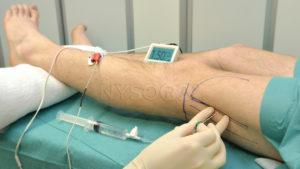
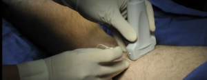
Popliteal Sciatic Nerve Block – Landmarks and Nerve Stimulator Technique
Features the technique description to perform a popliteal fossa block that results in anesthesia of the calf, tibia, fibula, ankle, and foot
Ultrasound-Guided Popliteal Sciatic Nerve Block Video
https://www.youtube.com/watch?v=gH1BS54EYjU