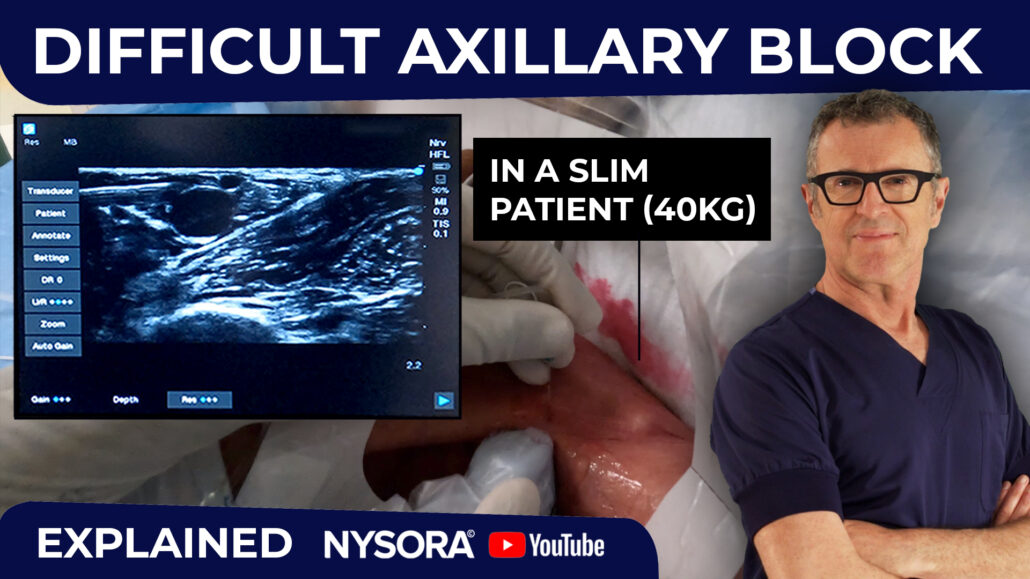
Axillary Brachial Plexus Block
Overview
- Indications: Forearm and hand surgery
- Landmarks: Axillary artery pulse
- Any of the following three endpoints
- Nerve stimulation: Hand twitch at 0.2-0.4 mA current
- Paresthesia: Hand
- Perivascular: Arterial blood aspiration (axillary artery)
- Local anesthetic: 35-40 mL
- Complexity level: Basic
|
|
General considerations
The axillary brachial plexus block was first described by Halstead in New York City at
St. Luke's-Roosevelt Hospital Center in 1884. The axillary brachial plexus block is a
basic nerve block technique, and one of the most commonly practiced blocks by
anesthesiologists in the United States. An axillary block is an excellent choice for
forearm and hand surgery. However, it should be noted that because the musculocutaneous
nerve leaves the brachial plexus sheath proximal to the site of injection, axillary brachial
plexus block often results in incon-sistent coverage for tourniquet pain as well as
anesthesia of the volar aspect of the skin below the elbow that extends to the thenar
eminence.
|
Regional anesthesia anatomy
The brachial plexus supplies innervation of the upper limb. The plexus consists of
nerves derived from the anterior rami of the lower four cervical and the first
thoracic spinal nerves. The five roots (anterior rami) give rise to three trunks
(superior, middle, and inferior) that emerge between the scalenus medius and scalenus
anterior to lie on the floor of the posterior triangle of the neck. The roots of the
plexus lie deep the prevertebral fascia. The trunks are covered by its lateral extension,
the axillary sheath. Each trunk divides onto an anterior and a posterior division behind
the clavicle, at the apex of the axilla. Within the axilla, the processes combine to produce
the three cords, which are named the lateral, medial, and posterior, according to their
relationships to the axillary artery. Each cord ends near the lower border of pectoralis
minor by dividing into two terminal branches. Other branches of the plexus arise from the
neck and axilla, directly from the roots, trunks, and cords. The anterior divisions form
the lateral and medial cords, with branches that supply the flexor muscle of the arm,
forearm, and hand, and the skin overlying the flexor compartments. The three posterior
divisions unite to form the posterior cord. The cord branches supply the extensor
musculature of the shoulder, arm, and forearm, and the skin of the posterior surface
of the limb.
Musculocutaneous Nerve
The musculocutaneous nerve is a terminal branch of the lateral cord. It pierces the
coracobrachialis muscle and lies between biceps and brachialis, supplying both of these
muscles. The nerve continues distally as the lateral cutaneous nerve of the forearm, which
pierces the deep fascia between biceps and brachioradialis to lie superficially over the
cubital fossa.
Median Nerve
The median and ulnar nerves traverse the entire length of the arm, but neither gives any
branches above the elbow joint. The median nerve derives its fibers from the lateral and
medial cords. In the upper part of the arm, the nerve lies lateral to the brachial artery.
However, at the midarm level, it crosses anterior to the vessels and finally lies medial
to the brachial artery, a position in which it continuous its path through the cubital
fossa. The median nerve enters the forearm from the cubital fossa between the two heads
of the pronator teres. It crosses anterior to the ulnar artery and descends between the
superficial and deep flexors. At the wrist, the median nerve is remarkably superficial,
lying medial to the tendon of flexor carpi radialis and just deep to the palmaris
longus tendon.
Ulnar Nerve
The ulnar nerve is a terminal branch of the medial cord. Together with the medial
cutaneous nerve of the forearm, the nerve initially lies medial to the brachial artery
but leaves the artery at midarm through the intermuscular septum. The nerve enters the
posterior compartment to lie between the septum and the medial head of triceps. The
ulnar nerve passes behind the medial epicondyle and enters the forearm between the two
heads of flexor carpi ulnaris. Lying on flexor digitorum profundus and covered by flexor
carpi ulnaris, it traverses the medial side of the anterior compartment, accompanied in
the lower part of the forearm by the ulnary artery. The ulnar nerve emerges near the
wrist lateral to the flexor carpi ulnaris tendon and crosses super-ficial to the flexor
retinaculum with the ulnar artery on its lateral side. The nerve terminates in the hand
by dividing into superficial and deep branches. The ulnar nerve supplies the elbow joint
and gives branches to the flexor carpi ulnaris and the medial part of flexor digitorum
profundus. It also provides a palmar cutaneous nerve that supplies the skin on the medial
aspect of the palm and dorsal cutaneous branches that innervate part of the medial part
of the dorsum of the hand.
Radial Nerve
The radial nerve, a terminal branch of the posterior cord, leaves the axilla by passing
below teres major and between the humerus and the long head of the triceps. The nerve
passes between the medial and lateral heads of triceps in the posterior compartment.
It then leaves the posterior compartment by piercing the lateral intermuscular septum
to reach the lateral part of the cubital fossa in front of the elbow joint. In the arm,
the radial nerve gives muscular branches to the medial and lateral heads of triceps and
branchioradialis and extensor radialis longus. Cutaneous branches innervate the lateral
aspect of the arm and the posterior aspect if the forearm. The branch to the long head
of triceps usually arises in the axilla.
Distribution of anesthesia
An axillary brachial plexus block (including musculocutaneous nerve block) provides
anesthesia to the to the arm, elbow, forearm, and hand. It should be noted that the
unshaded areas are not covered by the axillary brachial plexus block.
Patient positioning
The patient is in the supine position with the head facing away from the side to be
blocked. The arm on the side of the block placement should be abducted and form a roughly
90o angle in the elbow joint.
TIPS:
- Excessive abduction in the shoulder joint should be avoided because it makes
palpation of the axillary artery pulse difficult.
- Excessive abduction can also result in stretching and "fixing" of the brachial
plexus. Such stretching of the brachial plexus components increases the vulnerability
of the plexus during needle advancement. Stretching may increase the risk of nerve
injury because the plexus components are fixed and more likely to be penetrated by
the needle rather than "roll" away from the advancing needle.
Equipment
A standard regional anesthesia tray is prepared with the following equipment:
- Sterile towels and 4"x4" gauze packs
- 20-mL syringes with local anesthetic
- Sterile gloves, marking pen, and surface electrode
- 1½" 25-gauge needle for skin infiltration
- 3-5 cm long, short bevel, insulated stimulating needle
- A three-way stopcock
- Peripheral nerve stimulator
Landmarks
Surface landmarks
Surface landmarks for the axillary brachial plexus block include:
- Pulse of the axillary artery
- Coracobrachialis muscle
- Pectoralis major muscle
TIP: In some patients, palpation of the axillary artery may prove difficult;
a common scenario in young, athletic men. In this case, the approximate location of the
brachial plexus can be estimated by percutaneous nerve stimulation. The nerve
stimulator is set to deliver 4-5 mA and a blunt probe or an "alligator" clip
is firmly applied over the skin in front of the palpating fingers until twitches
of the brachial plexus are elicited.
Anatomic landmarks
After a thorough skin preparation, the pulse of the axillary artery is palpated high
in the axilla. Once the pulse is felt, it should be straddled between the index and
the middle finger and firmly pressed against the humerus to prevent "rolling" of the
axillary artery during block performance. At this point, movement of the palpating hand
and the patient's arm should be minimized because the axillary artery is highly movable
in the adipose tissue of the axillary fossa.
TIPS:
- When the location of the artery and the plexus is not immediately apparent,
asking the patient to adduct the arm against resistance during palpation of the
artery tenses the pectoralis and coracobrachialis muscles.
- This maneuver is helpful to identify the groove between coracobrachialis and
pectoralis muscle where the arterial pulse is easily detected.
- The position of the brachial plexus can be also estimated using percutaneous nerve
stimulation with a current output of 4-5 mA. A blunt probe or an "alligator" clip is
applied over the skin in front of the palpating fingers until twitches of the
brachial plexus are elicited. At this point, the probe is substituted by a needle
directed toward the estimated direction of the brachial plexus sheath.
Technique
After cleaning the skin with an antiseptic solution, local anesthetic is infiltrated
subcutaneously at the determined needle insertion site. The anesthesiologist
should assume a sitting position by the patient's side. This avoids strain and hand
movement during block performance and facilitates axillary block placement.
TIPS:
- Local anesthetic is best infiltrated tangentially rather than at a single
insertion point. This both ensures a superficial injection and allows for
needle repositioning during block performance if required.
- Hand position: The index and middle fingers of the palpating hand should
be firmly pressed against the arm, straddling the pulse of the axillary artery at
the midaxillary fossa level. This maneuver shortens the distance between the needle
insertion site and the brachial plexus block by compressing the subcutaneous
tissue. Also, it helps to stabilize the position of the artery and needle during
performance of the block. This hand should not be moved during the entire block
placement procedure to allow for precise redirection of the angle of the needle
insertion when necessary
Needle advancement
A needle connected to the nerve stimulator is inserted just in front of the palpating
fingers and advanced at an angle 45o cephalad. The nerve stimulator should be initially
set to deliver l mA current. The needle is advanced slowly until stimulation of the
brachial plexus, arterial blood, or paresthesia is obtained. Typically, this occurs
at a depth of 1-2 cm in most patients. Once the sought response is obtained, 35-40 mL
of local anesthetic is injected slowly with intermittent aspiration to rule out an
intravascular injection.
TIP: It has been suggested that the axillary brachial plexus sheath contains
septae preventing local anesthetic from reaching all neuronal components contained
within the sheath. While the clinical significance of these septae remains
controversial, it does make sense to inject local anesthetic in divided doses at
two different locations within the sheath (e.g., behind in front of the artery).
Goal
We use a nerve stimulator technique and look for a single nerve response, (hand twitch at
0.2-0.4 mA current output). We then inject the entire volume of local anesthetic on
obtaining such a response. Although multiple stimulation techniques (stimulating and
injecting each major nerve of the brachial plexus separately) may increase the success
rate, it also increases the complexity and time required to complete the block. However,
when the axillary artery is punctured before the plexus is stimulated (rare), we do not
continue searching for stimulation but resort to the transarterial technique and inject
one third of the total volume of the local anesthetic posterior and one third anterior
to the axillary artery.
Failure to obtain axillary brachial plexus stimulation on the first needle pass
When insertion of the needle does not result in nerve stimulations, the following maneuvers
should be made:
- Keep the palpating hand in the same position and the skin between the fingers stretched.
- Withdraw the needle to the skin, redirect the needle to angles of 15o
and 30o laterally and repeat the procedure.
- Withdraw the needle to the skin, redirect the needle to angles of 15o
and 30o medially and repeat the procedure.
Musculocutaneous nerve block
|
The musculocutaneous nerve is not consistently blocked with the axillary brachial plexus
block, because this nerve leaves the brachial plexus sheath proximally. Due to the large
area covered by this nerve and its importance in achieving complete anesthesia of the
forearm and biceps (see description in this chapter), a block of the musculocutaneous nerve
is often necessary for complete anesthesia. This is achieved with a separate injection by
inserting the needle above the artery and toward the coracobrachial muscle. Nerve stimulation
is used to produce twitches of the musculocutaneous nerve (biceps twitch). When twitches
are observed 5 mL of local anesthetic is injected. |
|
Troubleshooting
| Response Obtained |
Interpretation |
Problem |
Action |
| Local twitch of the arm muscles |
Direct stimulation of the biceps or triceps muscles |
The needle is inserted in direction that is too superior or too inferior |
Withdraw the needle and redirect accordingly |
| The needle contacts bone at 2-3 cm depth; no twitches are seen |
The needle is stopped by the humerus |
The brachial plexus was missed |
Withdraw the needle to the skin and reinsert at an angle 15o-30o superiorly or inferiorly |
| Twitches of the hand |
Stimulation of the medianus, radialis, or ulnar nerves |
Correct needle position |
Accept and inject local anesthetic. |
| Arterial blood noticed in the tubing |
Puncture of the axillary artery |
Needle entered the lumen of the axillary artery |
Inject 2/3 of the local anesthetic posterior to the artery and 1/3 anterior to the artery |
| Paresthesia- no motor response |
Contact of the needle with the brachial plexus branches |
Equipment malfunction (stimulator, needle, electrode) |
Carefully assess the distribution of the paresthesia and if typical, inject local anesthetic |
Choice of local anesthetic
The axillary brachial plexus requires a relatively large volume of local anesthetic (35-40 ml) to
achieve complete anesthesia. The choice of the type and concentration of local anesthetic should be
based on whether the block is planned for surgical anesthesia or pain management. Due to the highly
vascular area and potential for inadvertent intravascular injection, the local anesthetic solution
should be injected slowly, with frequent aspiration.
|
Onset (min) |
Anesthesia (hrs) |
Analgesia (hrs) |
| 1.5% Mepivacaine (+ HCO3; + epinephrine) |
5-15 |
2.5-4 |
3-6 |
| 2% Lidocaine (+ HCO3 + epinephrine) |
5-15 |
3-6 |
5-8 |
| 0.5% Ropivacaine |
15-20 |
6-8 |
8-12 |
TIPS:
- Always assess the risk-benefit ratio of using large volumes and concentrations of long
acting local anesthetic for this block.
- We never use bupivacaine for this block due to its high cardio-toxicity profile and potential
for inadvertent intravascular injection with axillary block technique.
- Smaller volumes and concentrations can be used successfully for analgesia (e.g, 15-20mL)
Block Dynamics and Perioperative Management
The axillary brachial plexus block is associated with relatively minor patient discomfort. Intravenous
midazolam 1-2 mg with alfentanil 250 to 500 µg at the time of the needle insertion should produce
a comfortable and cooperative patient during nerve localization. The onset time for this block is
rather long (15-25 minutes). The first sign of the blockade is the loss of the coordination of the
arm and forearm muscles. This sign can be seen usually sooner than the onset of sensory or temperature
change. When this sign is present within 1-2 minutes after injection, it has a very high positive
predictive value for a pending successful brachial plexus blockade.
Complications and How to Avoid Them
| Infection |
A strict aseptic technique is used. |
| Hematoma |
- Avoid multiple needle insertions, particularly in anticoagulated patients.
- Keep a 5-minute steady pressure when the axillary artery is inadvertently punctured.
- When planning to use a continuous technique, use a single-shot needle to localize the brachial plexus in patients with difficult anatomy.
- The use of antiplatelet therapy is not a contraindication for this block in the absence of spontaneous bleeding.
|
| Vascular puncture |
Steady pressure of 5 minute duration should be maintained when the carotid artery is punctured. |
| Local anesthetic toxicity |
- Systemic toxicity most commonly occurs during or shortly after injection of local anesthetic. This is most commonly caused by an inadvertent intravascular injection or channeling of forcefully injected local anesthetic into small veins or lymphatic channels cut during needle manipulation.
- Large volumes of long acting anesthetic should be reconsidered in older and frail patients.
- Careful and frequent aspiration should be performed during the injection.
- Avoid forceful, fast injection of local anesthetic.
|
| Nerve injury |
- Never inject local anesthetic when pressure on injection is encountered.
- Local anesthetic should never be injected when a patient complains of severe pain or exhibits a withdrawal reaction on injection.
|
| Total spinal anesthesia |
- When stimulation is obtained with a current intensity of <0.2 mA, the needle should be pulled back to obtain the same response with the current > 0.2 mA.
- Never inject local anesthetic when high pressure on injection is encountered.
|
Bibliography
- Benhamou D: Axillary plexus block using multiple nerve stimulation: a European view. Reg Anesth Pain Med 2001; 26:495
- Bouaziz H, Narchi P, Mercier FJ, Labaille T, Zerrouk N, Girod J, Benhamou D: Comparison between conventional axillary block and a new approach at the midhumeral level. Anesth Analg 1997; 84:1058-62
- Carles M, Pulcini A, Macchi P, Duflos P, Raucoules-Aime M, Grimaud D: An evaluation of the brachial plexus block at the humeral canal using a neurostimulator (1417 patients): the efficacy, safety, and predictive criteria of failure. Anesth Analg 2001; 92:194
- De Jong RH : Axillary block of the brachial plexus. Anesthesiology 1961; 22:215-25
- Finucane BT, Yilling F: Safety of supplementing axillary brachial plexus blocks. Anesthesiology 1989; 70:401-3
- Hadzic A, Vloka JD, Kuroda MM, Koorn R, Birnbach DJ: The practice of peripheral nerve blocks in the United States: a national survey. Reg Anesth Pain Med 1998; 23:241
- Hepp M, King R: Transarterial technique is significantly slower than the peripheral nerve stimulator technique in achieving successful block. Reg Anesth Pain Med 2000; 25:660
- Horlocker TT, Kufner RP, Bishop AT, Maxson PM, Schroeder DR: The risk of persistent paresthesia is not increased with repeated axillary block. Anesth Analg 1999; 88:382
- www.nysora.com, October 8, 2001.
- Klaastad O, Smedby O, Thompson GE, Tillung T, Hol PK, Rotnes JS, Brodal P, Breivik H, Hetland KR, Fosse ET: Distribution of local anesthetic in axillary brachial plexus block: a clinical and magnetic resonance imaging study. Anesthesiology 2002; 96:1315-24
- Lanz E, Theiss D, Jankovic D: The extent of blockade following various techniques of brachial plexus block. Anesth Analg 1983; 62:55-8
- Mezzatesta JP, Scott DA, Schweitzer SA, Selander DE: Continuous axillary brachial plexus block for postoperative pain relief. Intermittent bolus versus continuous infusion. Reg Anesth. 1997;22:357-62
- Partridge BL, Katz J, Benirschke K: Functional anatomy of the brachial plexus sheath: implications for anesthesia. Anesthesiology 1987; 66:743-7
- Schroeder LE, Horlocker TT, Schroeder DR: The efficacy of axillary block for surgical procedures about the elbow. Anesth Analg 1996; 83:747
- Selander D: Axillary plexus block: paresthetic or perivascular. Anesthesiology 1987; 66:726
- Selander et al: Catheter Technique in Axillary Plexus Block. Acta Anaesthesiol Scand 1977; 21:324-9
- Stan TC, Krantz MA, Solomon DL, Poulos JG, Chaouki K: The incidence of neurovascular complications following axillary brachial plexus block using a transarterial approach. A prospective study of 1,000 consecutive patients. Reg Anesth 1995; 20:486
- Thompson GE, Rorie DK: Functional anatomy of the brachial plexus sheaths.Anesthesiology 1983; 59:117-22
- Winnie A: Plexus Anesthesia; Vol 1: Perivascular techniques of brachial plexus blocks. Philadelphia, WB Saunders Co, 1984
- Winnie AP, Radonjic R, Akkinemi SR, Durrani Z: Factors influencing the distribution of local anesthetics in the brachial plexus sheath. Anesth Analg 1979; 58:225-34
GO TO TOP
|