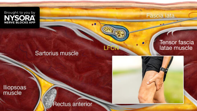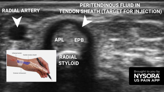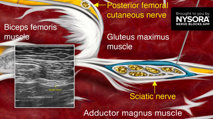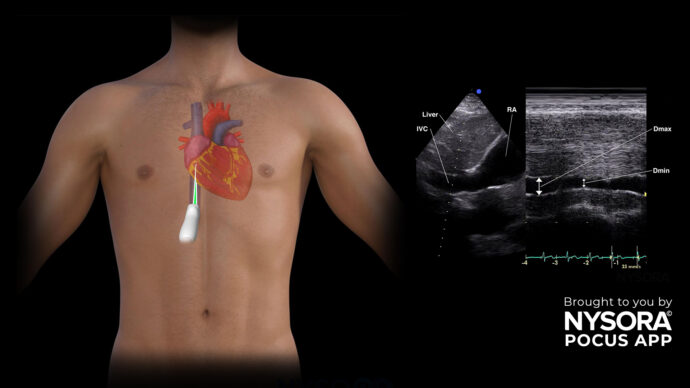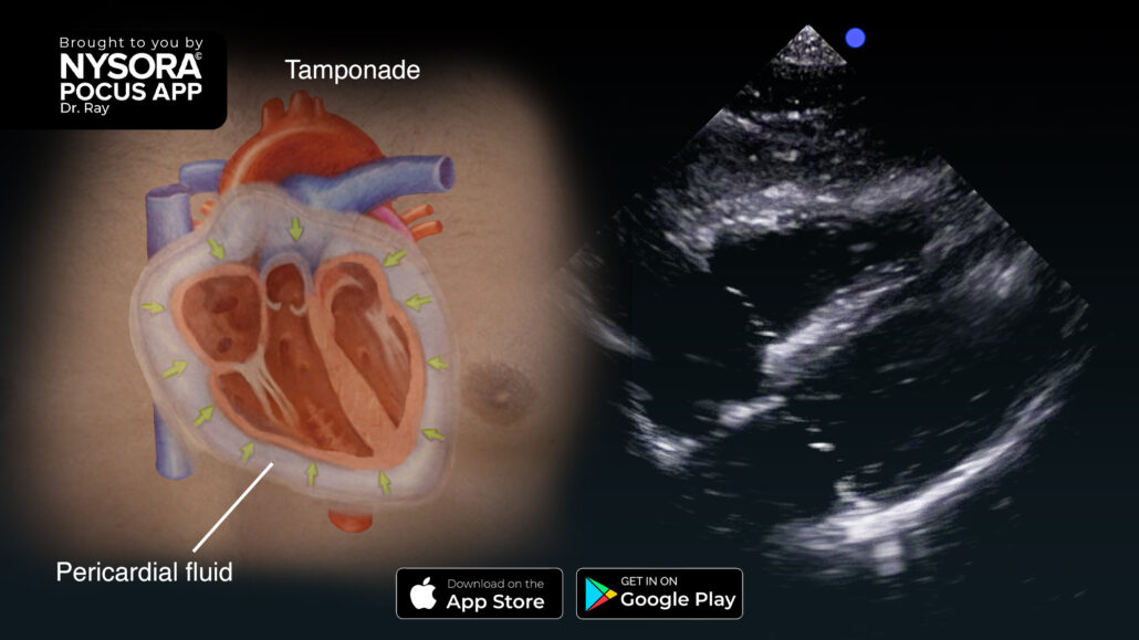
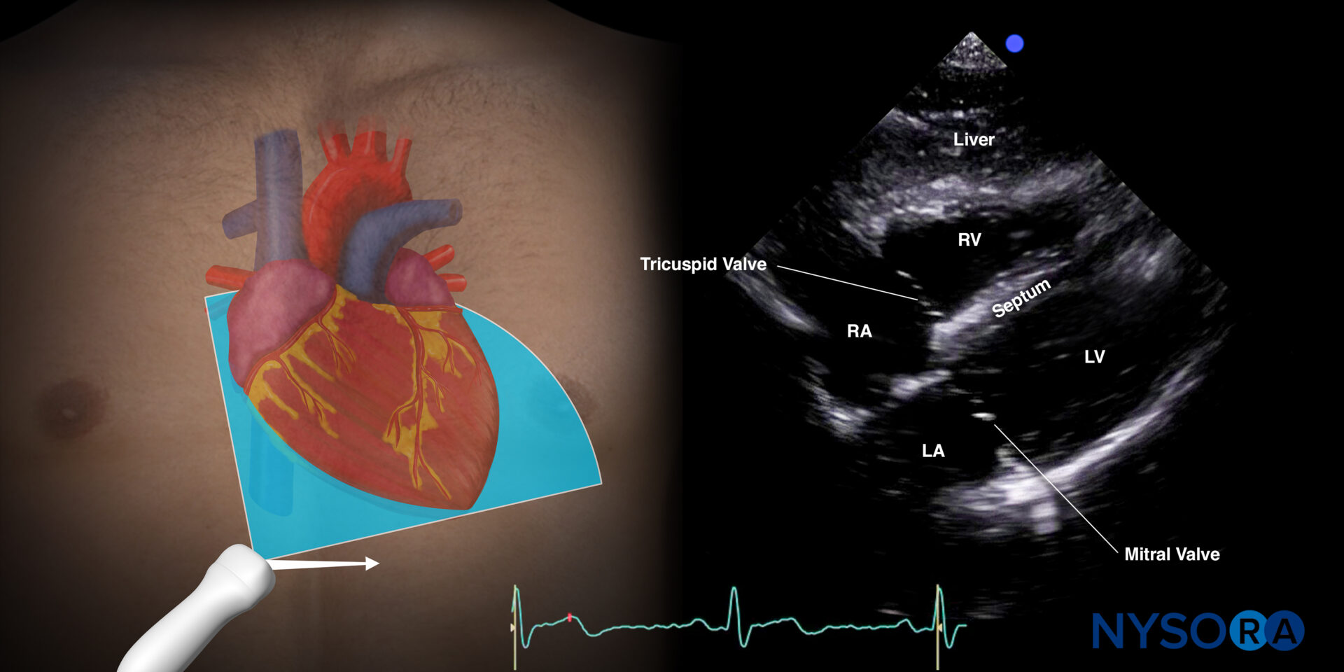
Identifying Pericardial Effusion or Tamponade: A Guide to Ultrasound Assessment in the Subcostal 4-chamber view (S4C)
The pericardium, a protective sac surrounding the heart, can accumulate fluid between its two layers (i.e., inner visceral and outer parietal pericardium) in various medical or surgical conditions. When this fluid buildup compresses the heart, leading to reduced cardiac output, it is known as pericardial effusion or tamponade. All views can be used to assess pericardial tamponade. However, did you know that the S4C is the best view to detect posterior collections?
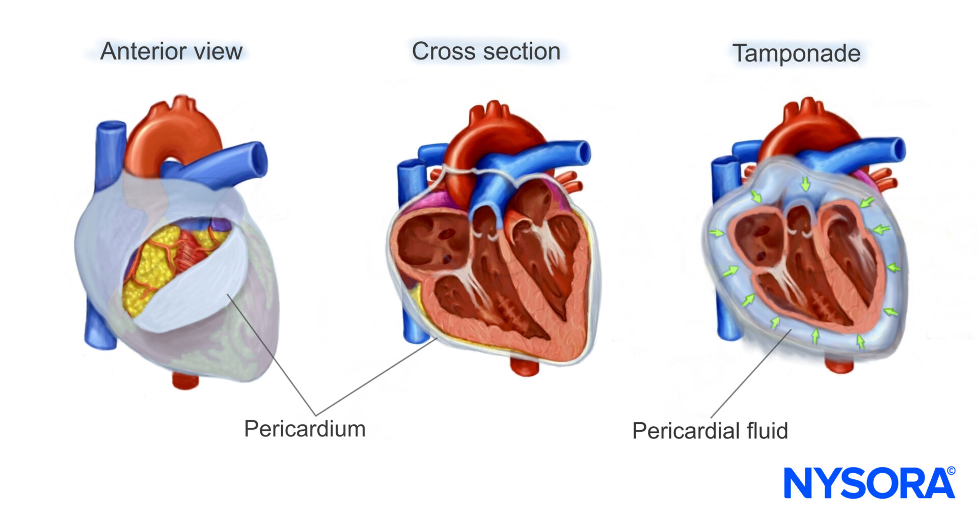
Ultrasound imaging plays a crucial role in identifying this condition and assessing its severity. Here’s a guide to detecting a large cardiac tamponade in the subcostal 4-chamber view (S4C).
Assessment
Ultrasound features: Anechogenic layer between the heart and pericardium. A large tamponade can be recognized by:
- Size: Measure at end-diastole! (overestimation during systole)
Large: > 2 cm - Cardiac cycle
During systole and diastole: Moderate – large - Location
Circumferential: Moderate – large - Additionally, you can visualize the inferior vena cava and focus on the right ventricle for compression.
Unleash the potential of POCUS with NYSORA’s POCUS App and elevate your practice, expand your capabilities, and deliver exceptional patient care.