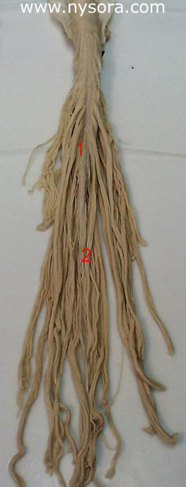
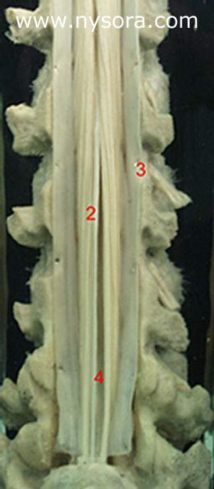
The lower extent of the spinal cord, the conus medullaris (1) ends in the adult at approximately L1. The line between the iliac crests, Tuffier’s line, most often crosses through the L4 spine. A line drawn between the posterior superior iliac spine identifies the level of the second sacral vertebras and the distant extent of the dural sac containing cerebrospinal fluid. The Fillum terminale (2) is a filament of connective tissue about 20 cm long, which descends from the apex of conus medullaris (1). Its upper 15 cm, the filum terminale internum, is surrounded by extensions of the dural (3) and arachnoid meninges and reaches the caudal border of the second sacral vertebra. This part is surrounded by a capacious space (4) filled with cerebrospinal fluid, and it is here where we perform spinal (lumbar) puncture and achieve spinal anesthesia by introducing local anesthetics. The final 5 cm of the filum terminale, the filum terminale externum, fuses with the investing dura mater, and then descends to the dorsum of the first coccygeal spinal nerves.
Anatomical material – courtesy of Prof. Dr. Faruk Dilberovic, Professor of Anatomy, Medical School Sarajevo, Bosnia and Herzegovina