Based on: Hadzic A, Vloka JD. A Comparison of the Posterior versus Lateral Approaches to the Block of the Sciatic Nerve in the Popliteal Fossa. Anesthesiology; 1988:88 (6):1480-1486. Vloka JD, Hadzic A, Kitain E, Lesser JB, Kuroda MM, April EW, Thys DM. Anatomic Considerations for Sciatic Nerve Block in the Popliteal Fossa Through the Lateral Approach. Reg Anesth 1996; 21:414-418.
|
Introduction The need to position the patient in the prone position is the main disadvantage of the posterior approach to the sciatic nerve block in the popliteal fossa (popliteal block; PB), and may preclude the use of the PB in patients who could benefit the most from this technique (i.e. advanced pregnancy, morbid obesity, spine and hemodynamic instability, mechanical ventilation). However, the lateral approach to PB results in reliable anesthesia, comparable to that of the PB using the posterior approach. The performance of the block using the lateral approach is straightforward when the described technique is followed, although it may take more attempts at nerve localization. In addition to using the PB in patients who can not assume the prone position, this technique provides the option of performing supplementary blocks (i.e., saphenous or femoral nerve blocks) and surgery without the need for patient repositioning. Technique The patients need adequate premedication (e.g., midazolam 10 to 30 µcg/kg and fentanyl 1 to 2 µcg/kg intravenously) prior to introduction of the anesthetic. Additional doses of midazolam and fentanyl in increments of 1 mg and 50 µcg, respectively, are administered as necessary during the block performance and surgery, in order to allay any accompanying anxiety and discomfort.
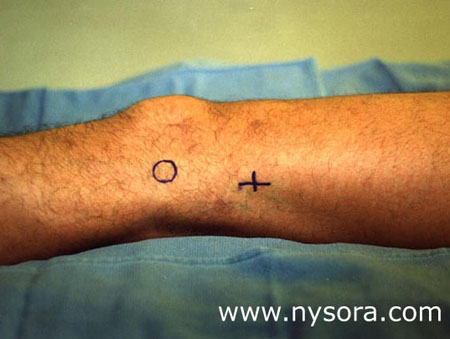
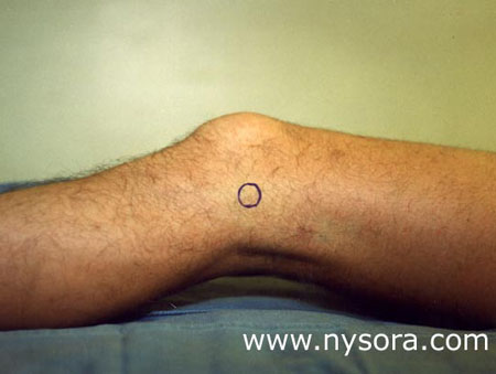
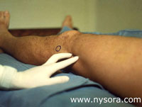
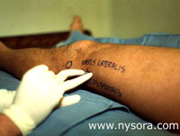
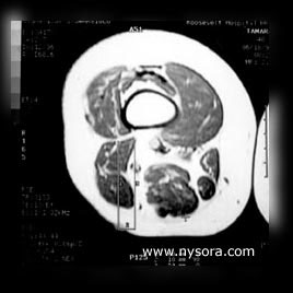
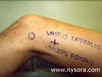
The skin was prepared with a solution of povidone iodine, and the site of needle insertion was infiltrated with 2 mL of 1% lidocaine using a 25 gauge needle. The lateral approach to PB is performed with patients in the supine position and with their legs extended in the knee joint (Figure 1). The long axis of the foot is positioned at a 90 degree angle to the table. A 100 mm long, 21G insulated stimulating needle attached to a nerve stimulator (Stimuplex®, B. Braun Medical, Inc., Bethlehem, PA), attached to a nerve stimulator is inserted in a horizontal plane 7 cm cephalad to the most prominent point of the lateral femoral epicondyle, in the groove between the biceps femoris and the vastus lateralis muscles (Figures 2, 3, and 4) until the shaft of the femoral bone is intentionally contacted. When the femur is not contacted within the depth of approximately 50 mm, the needle is reinserted 5 to 10 mm anteriorly (above) to the first insertion.
After the femur is contacted, needle is withdrawn to the skin, and redirected posteriorly at a 30 degree angle to the horizontal plane (Figure 5): 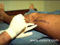 Figure 5. After the femur is contacted, the needle is withdrawn to the skin and redirected posteriorly at an angle of 30 degrees to the one at which the femur was contacted.
The initial current of 0.8 mA (1 Hz) is gradually decreased in both approaches after obtaining an initial response to nerve stimulation. The localization of the nerve is considered successful when either tibial nerve response (plantar flexion), or common peroneal response (lateral inversion-dorsal flexion) is obtained. The output current of the nerve stimulator is adjusted to the lowest current at which these responses are still observed. At this point, after inadvertent intravascular placement of the needle is ruled out by gentle aspiration, 40 mL of 1.5% alkalinized mepivacaine (1 mEq of NaHCO3 per 30 mL of mepivacaine) with 1:200,000 epinephrine is injected. When stimulation of the nerve required a a current of greater intensity the local anesthetic solution was injected only after the response of thcurrent greater than 0.4 mA, an attempt is made to stimulate the division of the popliteal nerve that predominantly innervated the surgical area. When necessary, a supplementary block of the saphenous nerve is performed at the level of the tibial tuberosity using 8 to 10 mL of pH-adjusted 1.5% mepivacaine with 1:200,000 epinephrine. If the stimulation of the sciatic nerve is not obtained, the needle is withdrawn to the skin and reinserted through the same skin puncture, first 5 to 10 degrees anterior and then 5 to 10 degrees posterior relative to the initial insertion (30 degree) plane. If this redirections (1st attempt) do not result in nerve localization, the same technique is repeated through new skin punctures (subsequent attempts) in 5 mm increments posterior to the initial insertion plane.
|