Clinical case: Left ventricular function
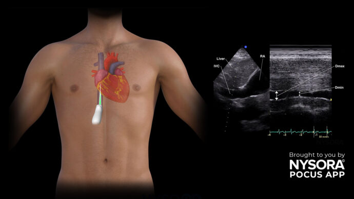
A 71-year-old male with a medical history of coronary artery disease, chronic hypertension, and type 2 diabetes presents with chest pain. Upon arrival, standard protocol led to the execution of an ECG, chest X-ray, and blood tests. While awaiting these results and during the clinical examination, a point-of-care ultrasound scan focusing on the heart was conducted, comprising five crucial ultrasound views. Each view can be used to evaluate the left ventricular function, and, in this case, the apical four-chamber view was used.
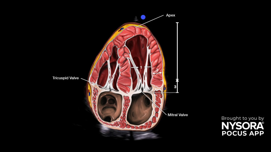
The technique, also known as eyeballing, evaluates the inward motion of the endocardium, the thickening of the left ventricular wall during systole, and the longitudinal shortening or movement of the mitral valve apparatus toward the heart’s apex. Closely assessing these 3 signs in the apical four-chamber view can distinguish a normal from a decreased left ventricular function.
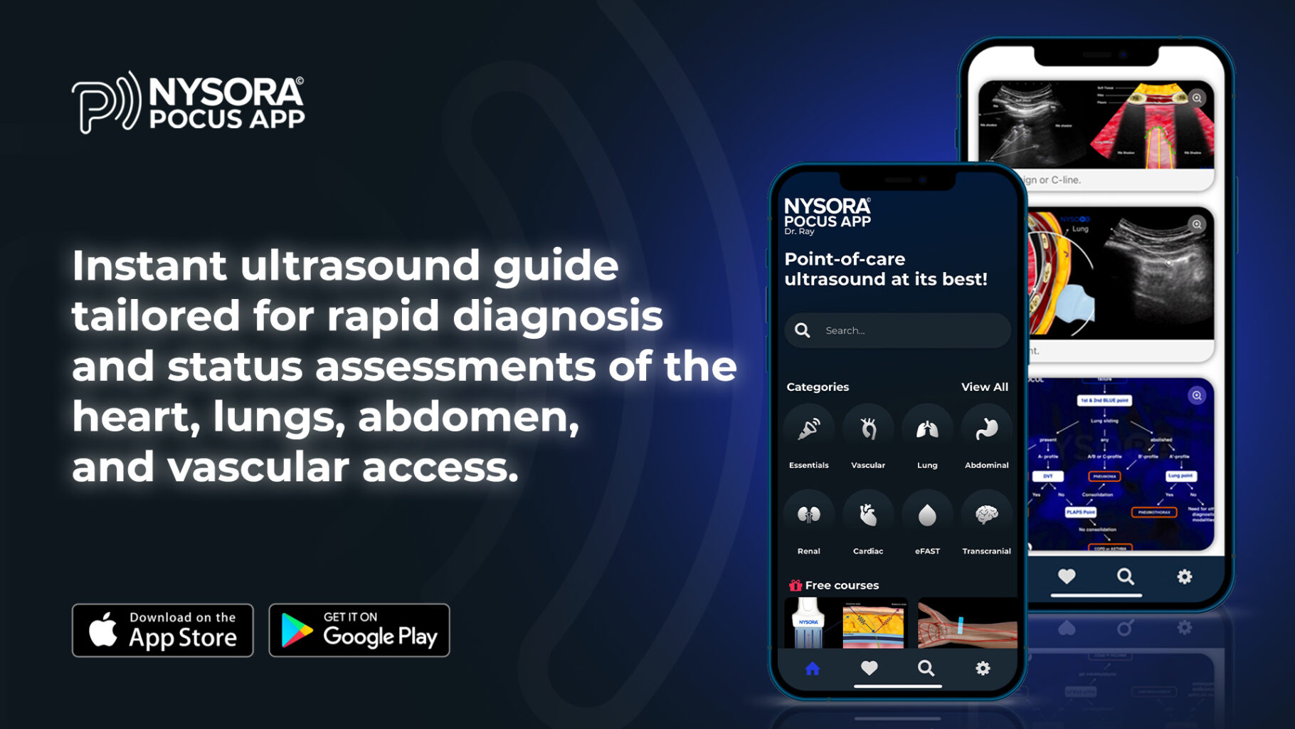
Unleash the potential of POCUS with NYSORA’s POCUS App and elevate your practice, expand your capabilities, and deliver exceptional patient care. Download HERE.